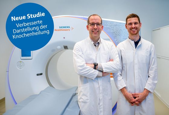
A pioneering study has shown that the visualization of bone healing in patients with metal implants can be significantly improved by the reduction of implant artifacts. The study was conducted by Dr. Adrian Marth, radiologist at the Swiss Center for Musculoskeletal Imaging at Balgrist Campus and led by Prof. Reto Sutter, Head of Radiology at Balgrist University Hospital. The researchers investigated the clinical benefit of the combination of tin prefiltration and virtual monoenergetic image reconstructions (VMIs) in CT examinations with a novel photon counting detector (PCD) CT system.
The study was conducted last year in 48 patients with metal implants in the feet or lower leg. VMIs with different energy levels between 60 kilo-electronvolts (keV) and 190 keV were created from the spectral image data to determine the optimal energy to reduce implant artifacts and improve the visibility of bone healing. The study demonstrated a significant advantage of CT imaging with tin prefiltration and VMIs at an energy level of 120 keV, which substantially improves the visualization of bone healing.
"To my knowledge, this is the first scientific study worldwide to show that the higher spatial resolution of photon counting CT has a clinical impact in the assessment of bone healing in patients. The significant reduction of metal implant artifacts due to tin prefiltration and monoenergetic image reconstruction and thus an overall higher image quality improves the visualization of bone healing. This is important for assessing the healing process in patients with metal implants," says Reto Sutter, Head of Radiology at Balgrist University Hospital.
Reference
Marth AA, Goller SS, Kajdi GW, Marcus RP, Sutter R. Photon-Counting Detector CT: Clinical Utility of Virtual Monoenergetic Imaging Combined With Tin Prefiltration to Reduce Metal Artifacts in the Postoperative Ankle. Investigative Radiology 2024 online before print
For further information please contact
Gregor Lüthy, Head of Communications
Balgrist University Hospital
+41 44 386 14 15
Email

