Radiological diagnostic investigation of the spine
Radiology at the University Spine Center in Zurich is highly specialized and concentrates on the diagnosis of spine diseases. Investigation of the spine is a focal point of our clinical and scientific work.
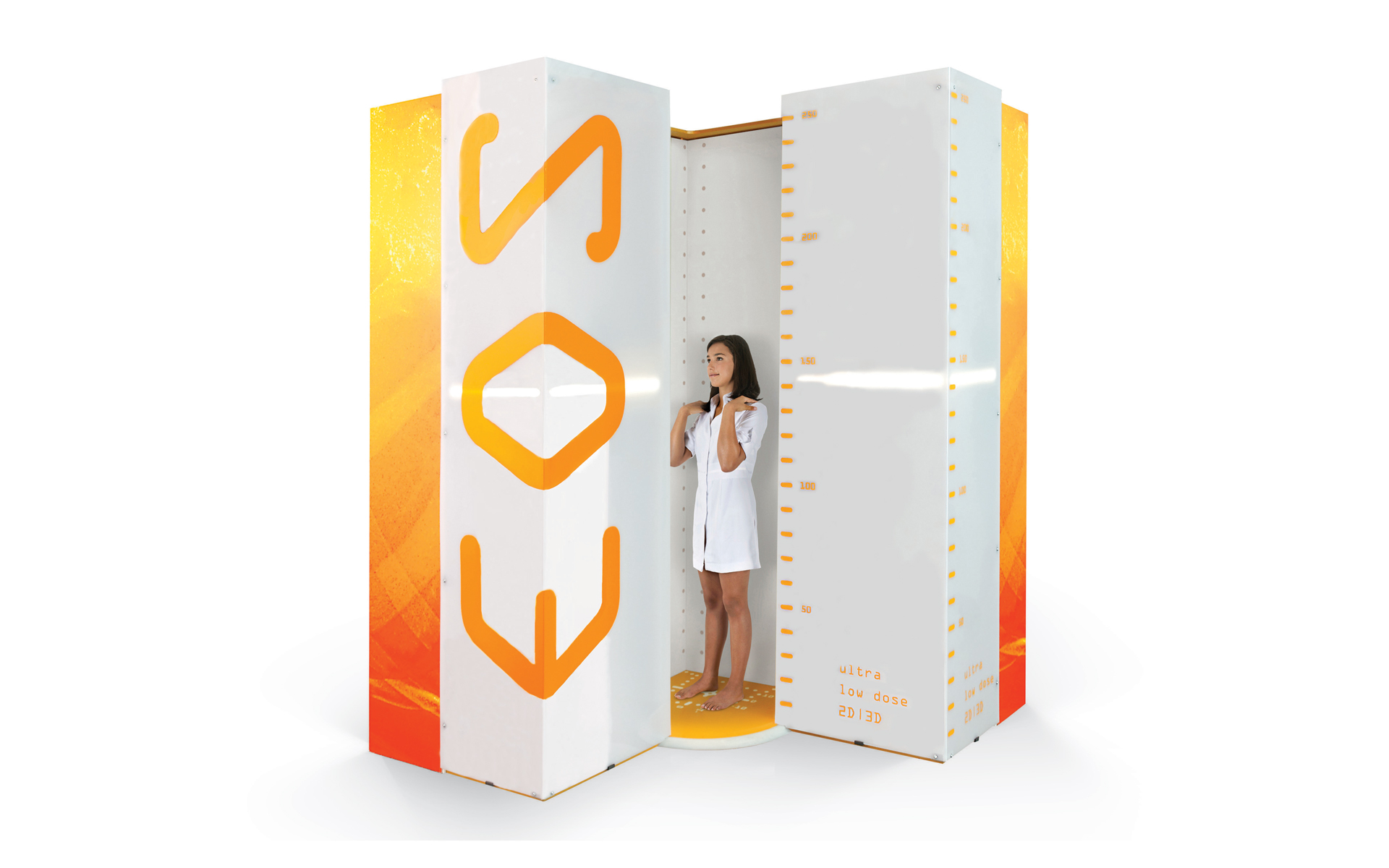
State-of-the-art EOS scanner
EOS imaging is a state-of-the-art procedure used especially for examination of the spine. The patient stands in a comfortable position for scans in two planes. The radiation exposure is much less than with normal X-rays. The whole spine or even the whole body can be scanned to assess the static structures. 3D analysis allows us to take precise measurements.
Greatly reduced radiation exposure
In ‘micro-dose’ mode, we can reduce the radiation even further to an extremely low dose. Your exposure is then roughly the same as a few weeks natural background radiation. The micro-dose examination is particularly suitable for the analysis of deformities in which the details of the bone structure do not have to be analyzed.
Minimally invasive treatment of spinal pain
The University Spine Center has a decade of experience of minimally invasive treatment for back pain. Image-guided minimally invasive therapy is targeted on pain starting in the spine. Painkillers and anti-inflammatory drugs are injected around irritated nerve roots close to a prolapsed disc or into the small joints of the spine. Using CT for guidance allows us to insert the needle gently and with millimeter precision. We use the latest equipment with low radiation exposure. Ultra-low-dose protocols are used for image-guided minimally invasive treatment. In addition, we offer the possibility of pulsed radiofrequency therapy to treat facet joint arthritis and nerve root irritation. The quality of minimally invasive pain therapy of the spine is continuously scientifically scrutinized and improved.
Bone quality
Assessing the quality of bone is an integral part of our investigations at the University Spine Center. Spinal bone quality is of key importance for stability. Fractures – due to osteoporosis – are serious events that cause pain and changes in the statics of the spine. Bone quality is assessed with bone densitometry (DEXA).
The spine after an operation
Investigation of the spine after an operation is often particularly challenging. Implants cause artefacts in CT and MRI scans. Research in the radiology department of the University Spine Center has made a significant contribution to reducing these artefacts and to improving the quality and informative value of the investigations. We use protocols adjusted specifically to the postoperative situation.
Here for you

Prof. Dr. med. Reto Sutter
Head Physician Radiology
Appointments
+41 44 386 16 00
Email
Further information
The radiology team led by Prof. Dr. med. Reto Sutter carries out diagnostic imaging of the musculoskeletal system using a whole range of state-of-the-art X-ray, fluoroscopy, ultrasound, CT, and MRI equipment. The team focusses entirely on the musculoskeletal system, making it uniquely experienced in this field in Switzerland. The radiology services are directly available for all our patients and for referrals from other doctors and hospitals.

